Search
Croatia
Electro Cardio Graph (ECG) 
Remote Patient Monitoring System
Anatomy of the Heart

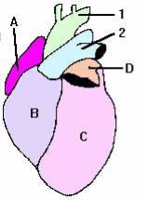
Figure 1. The main parts of the heart

The heart supplies the body tissues with oxygen saturated blood, it also ensures central heating and cooling and a number of other vital functions. If the heart fails to function death occurs within minutes.

The heart consists of muscle tissue and, as a contrast to most other organs in the body, it generates its own electrical impulses ensuring automatic function; pumping blood.


The heart has the following main parts:

A - Right ventricle

B - Left ventricle

C -  Right atrium

D - Left atrium

1 - Aorta

2 - Pulmonary artery


Figure 2. The electrical pathway of the heart.


The electrical signals which produce the pumping of the atria and the ventricles is generated in two nodes (see figure 2); the SA node for the atria (starting each sequence) and the AV node for the ventricles. The signal from the SA node depolarizes the atria and elicits the AV node to fire its impulse. This, turn depolarizes the ventricles which pump containing blood through the aorta into the body as well as blood to the lungs for “oxygen-filling”.


When measuring the ECG electrodes are placed on the skin of the patient. A one lead-system only measures the ECG at one point. By using 3, 5 or 12 leads you improve the ECG resolution enable interpretation of a number of diseases and/or complications (e.g. Cardiac infarction). The beneficial effect is achieved by reading electric signals from different angles and distances from the heart. In the most advanced setting, 12 lead ECG electrodes are placed on the chest (see figure 3) and on each arm and leg.

Figure 3.
Figure Description

Electrode placement on the chest

Normal sinus rhythm

Normal sinus rhythm is the rhythm of a healthy normal heart, where the sinus node triggers the cardiac activation. The sinus rhythm is normal if its frequency is between 60 and 100/min (See figure 4). If the sinus rhythm frequency is less than 60 it is called Bradycardia and if it is above 100 it is called Tachycardia.

Figure Description

A normal ECG curve in Sinus Rhythm with the electrical signal starting from the S-A Node

Atrial Flutter

When the electric signals fire too rapidly the pumping effect of the heart atria’s is replaced by ineffective atrial flutter. The frequency of the contractions is between 220 and 300/min. The ventricles are generally activated by every second or every third atrial impulse (2:1 or 3:1 heart block).


A patient may live with atrial flutter but feels very weak and short of breath during episodes. Drugs may cure the symptom and stabilize the signals. Patients having chronic symptoms normally gets a pace maker.

Figure Description

Atrial Flutter, rapid flutter waves. Ventricular response irregular.

Ventricular Flutter

When ventricular depolarization occurs chaotically, the situation is called ventricular fibrillation. The lack of blood circulation leads to almost immediate loss of consciousness and death within minutes. The ventricular fibrillation may be stopped with an external defibrillator pulse and appropriate medication.

Figure Description

Ventricular Flutter, rapid wide irregular ventricular complexes.